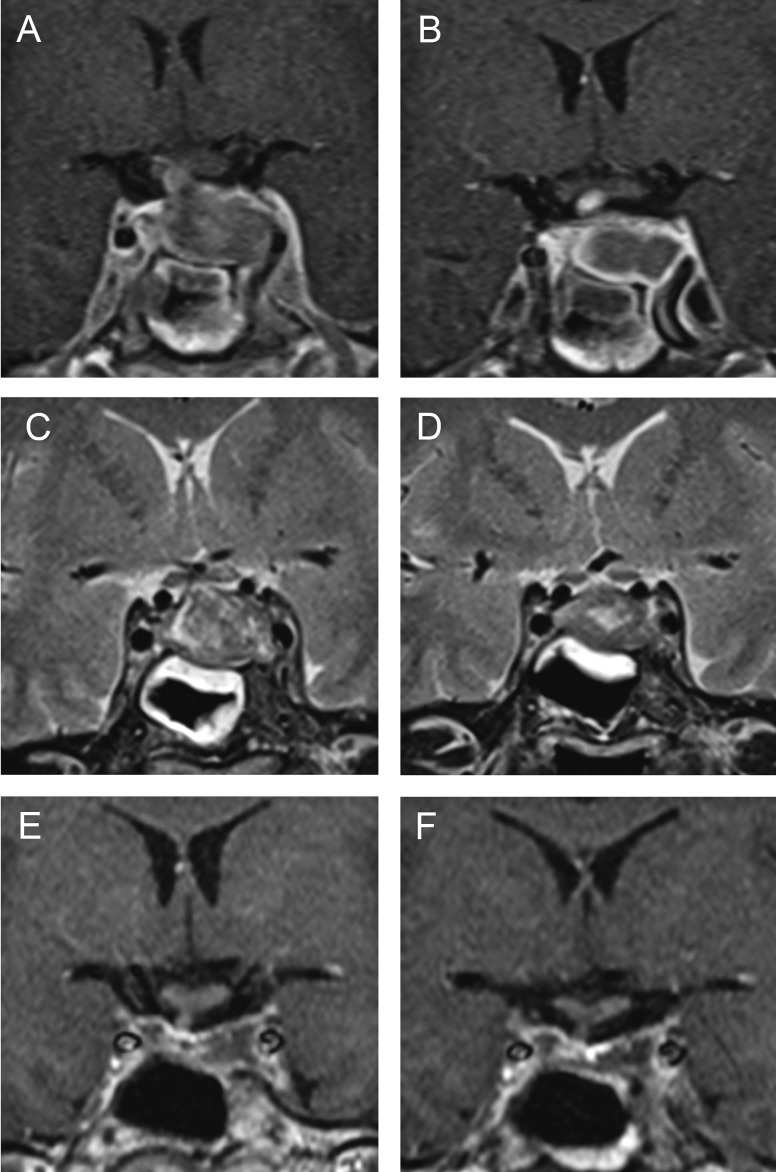
Fig. 1.
Magnetic resonance imaging in a patient with necrotizing hypophysitis showing lack of enhancement within enlarged pituitary gland on contrast-enhanced imaging, which is hypointense to isointense to the gray matter on T1-weighted images and thickened pituitary stalk (A); progression of ischemia 12 days after symptom onset with rim enhancement on T1-weighted images (B); thickened sphenoidal sinus mucosa on T2-weighted images (C), and regression of edema twelve days after symptom onset (D); residual necrotic pituitary tissue on contrast-enhanced imaging 4 (E) and 18 (F) months after the surgery.

