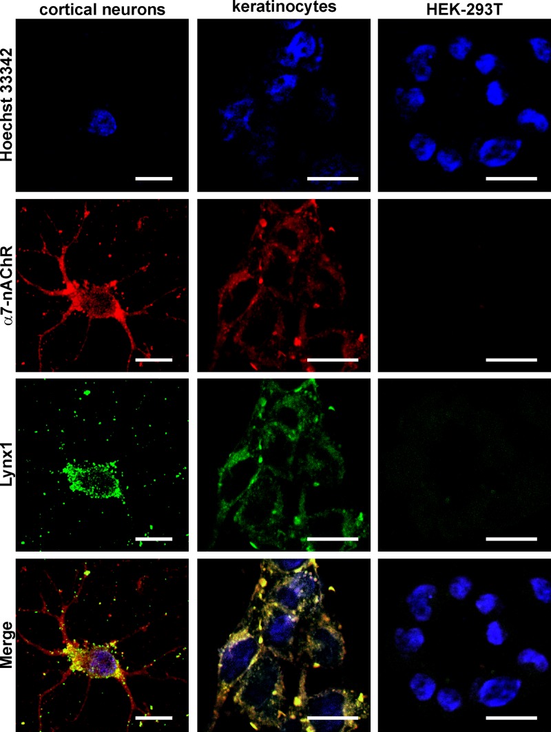
Fig 2. Colocalization of endogenous Lynx1 and α7-nAChRs in primary cortical neurons, oral Het-1A keratinocytes and HEK-293T cells.
Cells were sequentially incubated with the rabbit anti-Lynx1 and mouse anti-α7-nAChR primary antibodies and with secondary anti-rabbit Alexa-488 labeled IgG (green) and anti-mouse Alexa-594 labeled antibodies (red). Cell nuclei were visualized by Hoechst 33342 (blue). Scale bar 10 μm.

