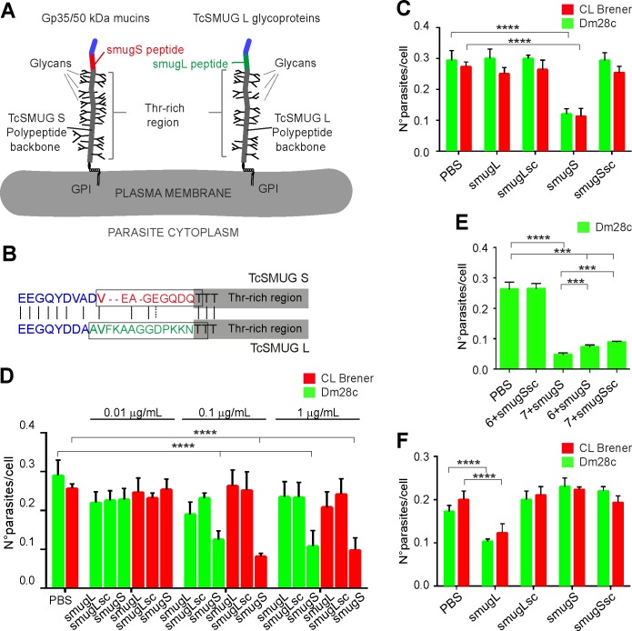
Fig 5. Peptidic determinants mediate adhesion of Gp35/50 kDa mucins to T. infestans rectal ampoule.
A) Schematic diagram showing the structural features and topological disposition of surface-displayed Gp35/50 kDa mucins and TcSMUG L glycoproteins. The relative position of the smugS and smugL peptides on the overall polypeptide is indicated. GPI, glycosylphosphatidyl inositol. B) Sequence alignment between the mature N-terminal regions from TcSMUG S and TcSMUG L canonical proteins. Amino acids are colorized in accordance to their relative position on the polypeptide, as defined in A. Residues included in the smugS and smugL peptides are boxed. C-F) Freshly dissected rectal ampoules (C-E) or midgut tissues (F) obtained from fifth-instar nymphs 12–15 days after the last bloodmeal were incubated for 30 min in PBS or PBS supplemented with the indicated synthetic peptide (at variable concentrations in panel D; at 0.1 μg/mL in panels C and F and added with interaction medium containing 2 x 10³ CL Brener or Dm28c epimastigotes. In panel E, tissues were incubated for 30 min in PBS or PBS supplemented with the indicated synthetic peptide (0.1 μg/mL) and carbohydrate compound (as numbered in Fig 4A; 20 nM each). In all cases, adhered epimastigotes were counted per 100 epithelial cells in 10 different fields of each hindgut preparation. For each experimental group, 10 insects were used and experiments were performed in triplicate. Data are expressed as mean ± S.D. and asterisks denoted significant differences (*** when P < 0.001; and **** when P < 0.0001) between each of the indicated populations and the control (PBS treated) population, as assessed by ANOVA and Tukey's tests. In panel E, additional pairwise comparisons were carried out (compound 7 + smugS peptide vs compound 6 + smugS peptide and compound 7 + smugS peptide vs compound 7 + smugSsc peptide), and results are indicated as above.

