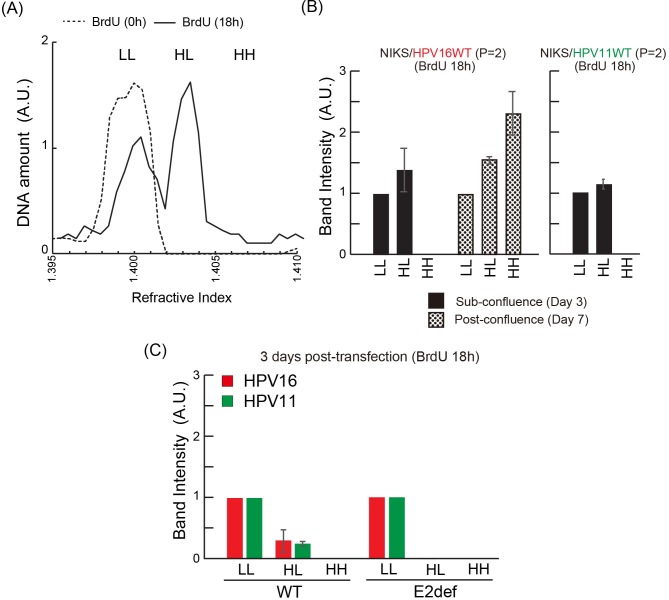
Fig 4. Synchronous HPV DNA replication durind sub-confluent growth.
(A) Profile of NIKS cellular DNA 18 h after BrdU labelling. The DNAs were separated on a CsCl gradient, and fractions were collected. In each fraction, refractive index (RI) and DNA concentration were measured by a spectrophotometer and a refractometer respectively. The positions of heavy-heavy DNA (HH), heavy-light DNA (HL), and light-light DNA (LL) are marked. (B) DNA of NIKS containing HPV16 or 11 was collected at day 3 and 7 of passage 2 following 18 h of BrdU labelling. The DNAs were separated on a CsCl gradient, and fractions were collected as before. Fractions where the RI indicated the possible presence of DNA (HH, HL or LL) were blotted, before the detection and quantitation of HPV DNA using DIG-labelled probes. (C) DNA from NIKS containing HPV16 or 11 genomes was collected at 3 days post-transfection following 18 h of BrdU labelling. HH, HL or LL HPV DNA was separated and detected as described in (B).

