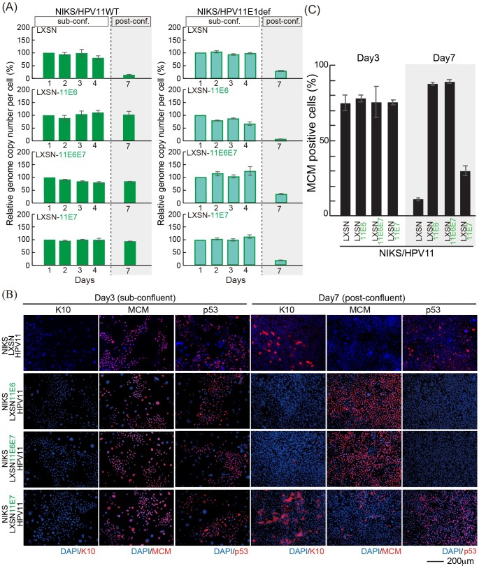
Fig 7. Low-risk HPV is maintained in NIKS expressing low-risk HPV E6 and E7 post-confluence.
(A) The virus genome copy number per cell of HPV11 (left) and HPV11 E1def (right) transfected into NIKS or NIKS expressing HPV11 E6 and/or E7 was measured and prepared for presentation as outlined in Fig 1E. (B) The NIKS and NIKS expressing HPV11 E6 and/or E7 at day 3 and 7 were stained with K10 (red), MCM (red), p53 (red) and DAPI (blue). Bar = 200μm. The images of NIKS at day 3 and 7 were shown previously in Fig 3C. (C) The proportion of MCM positive cells shown in (B) is quantified and shown.

