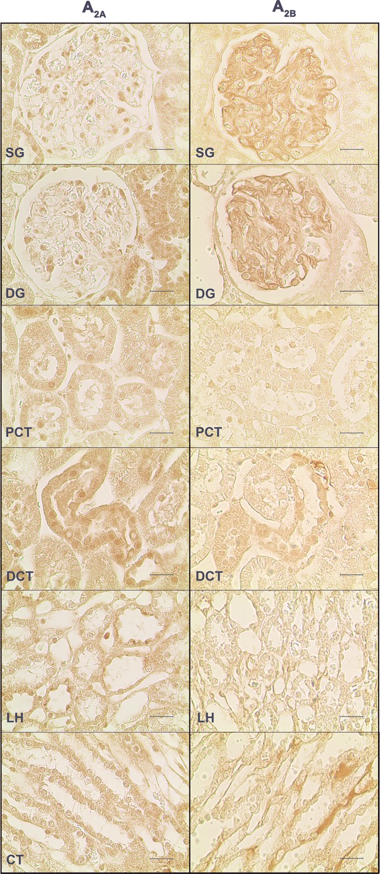
Fig 3. Immunoreactivity against the adenosine A2A and A2B receptors receptors in the SHR control group.

Representative photomicrographs of kidney sections from 4 SHR control rats incubated with a primary antibody against the adenosine A2A (left panels) and A2B (right panels) receptors. The six renal structures studied in separate are represented: superficial (SG) and deep glomeruli (DG), proximal (PCT) and distal (DCT) convoluted tubule, loop of Henle (LH) and collecting tubule (CT). Scale bars: 20 μm.
