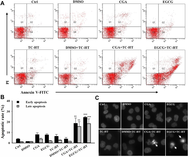
Fig 4. Combination of the TC-HT and the CGA or EGCG induces apoptosis in PANC-1 cells.
The apoptosis analysis for the cells following the treatment with the TC-HT (10 cycles), 0.05% DMSO (vehicle control), CGA (200 μM), and EGCG (20 μM) alone or in combination (TC-HT + DMSO, TC-HT + CGA, TC-HT + EGCG) for 24 h. (A) Flow cytometric detection of the apoptosis with Annexin V-FITC/PI double staining. (B) Histogram quantifying the percentage of PANC-1 cells in early and late apoptosis. (C) The nuclei morphology alterations (arrow) were examined using DAPI staining. Data are presented as mean ± S.D. in triplicate. (**p < 0.01 and ***p < 0.001 compared with the untreated control).

