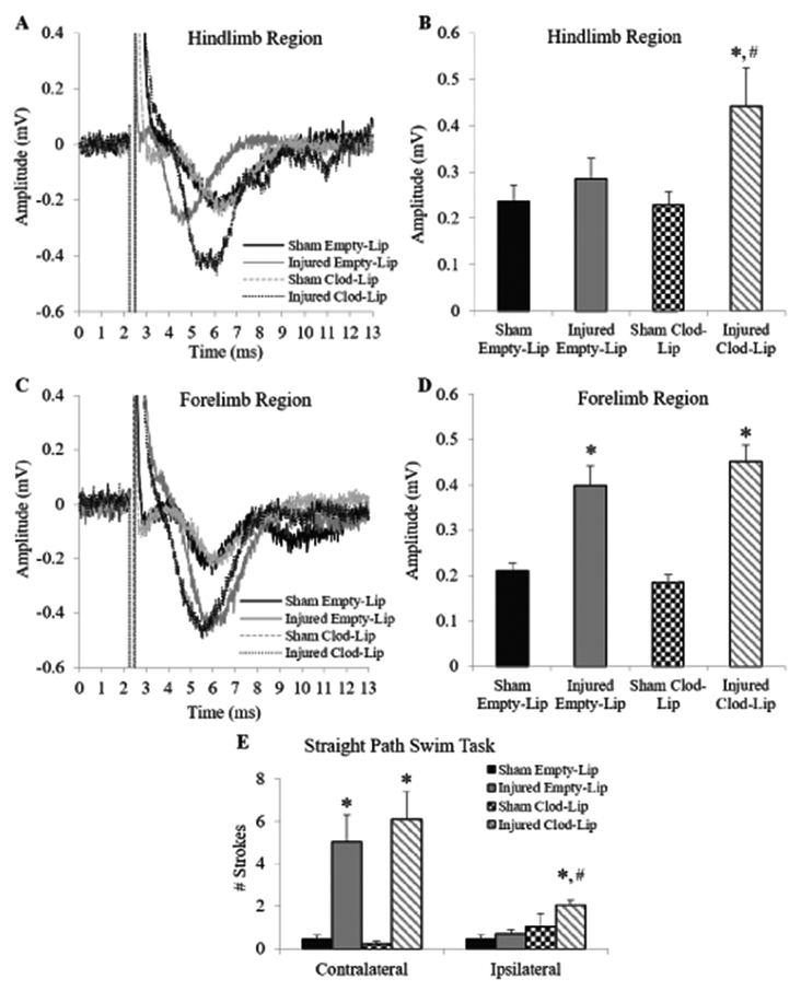
Figure 4. Extracellular evoked potentials in the cortex at 4 weeks following clodronate-induced microglia depletion.
Representative traces from the hindlimb region (A) and forelimb region (C) of the motor cortex of sham-injured and brain-injured animals that received either empty or clodronate liposomes. (B, D) Quantification of the amplitude of the maximum deflection of the traces from the respective regions. (E) Forelimb motor deficits in the straight path swim task. Bar graphs represent group means and error bars represent the standard error of the mean. *, p≤0.05 compared to corresponding sham-injured group; #, p≤0.05 compared to corresponding brain-injured animals injected with empty liposomes.

