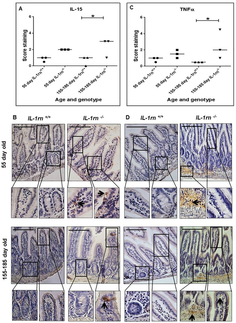
Figure 5.
Immunohistochemistry staining of the expression and localization of pro-inflammatory cytokines: IL-15 (A) TNFα (C) show the immunopositive intensity quantification across the small intestinal architecture. (B and D) showing immunopositive staining in the ileum of the 55 day old and 155-185 day old IL-1rn-/- mice compared with WT mice. Cell nuclei were stained with haematoxylin (blue). Black arrows indicate immunopositivity. *P ≤ 0.05. Scale bar = 100 µm.

