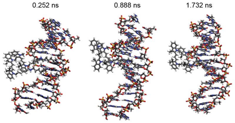
Figure 5.
Snapshots of MD simulations at the specified time frames. The Cy3 molecules are visible on the left and they are located, throughout the whole simulation, close to the DNA backbone. No intercalation is observed during the observation time of 2 ns. The molecules show a non-prefect parallel alignment and a fluctuating reciprocal position, which is consistent with the experimental observations of CD and the co-existence of monomer and dimer peaks.

