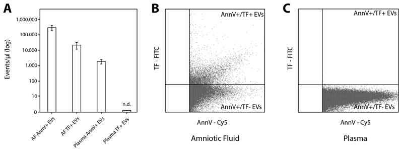
Figure 1. Extracellular vesicles (EVs) quantified by flow cytometry.
(A) PS+/Annexin V+ EVs were significantly higher in AF samples (n=10) compared to plasma samples from healthy controls (n=7) (A and B). In AF a high proportion of detected PS+ EVs also exposed TF (A and C). In the plasma of healthy controls no TF+ EVs were detectable. Single measurements were performed. AF, amniotic fluid; PS, phosphatidylserine; TF, tissue factor.

