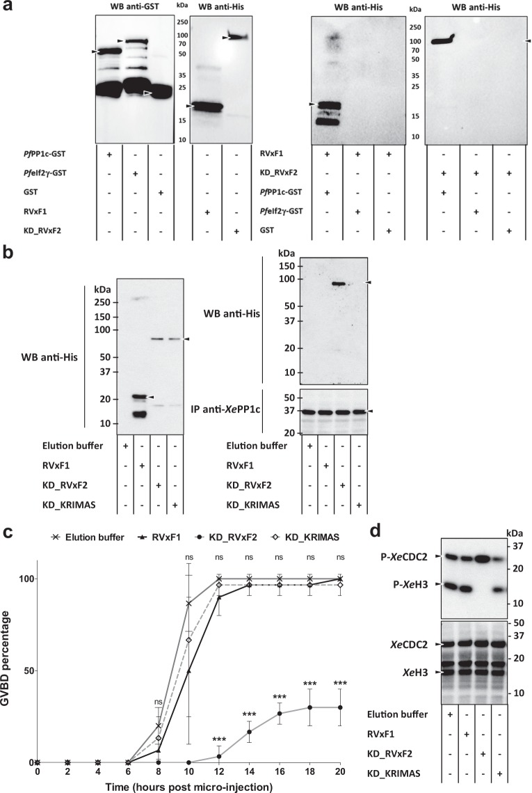
Figure 5.
Both PfpTKL RVxF1 and RVxF2 motifs bind PfPP1c in vitro but only RVxF2 exerts a functional role in Xenopus laevis oocytes. (a) RVxF1 and RVxF2 both directly bind PfPP1c in vitro. PfPP1c-GST, PfeIf2γ-GST or GST (the last two = negative control) beads were incubated with His6-pTKL_RVxF1 (RVxF1) or pTKL_KD_WT (KD_RVxF2). Following washes, beads were eluted in Laemmli buffer. Proteins were separated by SDS-PAGE and His6-tagged proteins bound to beads were identified by western blot (WB, right panel – both blot first lanes show the specific and direct interaction of RVxF1 and KD_RVxF2 with PfPP1c-GST). All inputs were analyzed by western blot as well (left panel). Uncropped blots are available in the Supplementary Information. (b) Only RVxF2 binds XePP1c in vivo. RVxF1, KD_RVxF2 and KD_KRIMAS (containing RVxF2 inactivated by mutation) were micro-injected into X. laevis oocytes. To check for the presence of RVxF-containing proteins, oocytes were lysed and His6-tagged proteins were separated by SDS-PAGE and detected by western blot (left panel). Oocytes micro-injected with RVxF1, KD_RVxF2 or KD_KRIMAS and incubated with progesterone were lysed 15 h after micro-injection and anti-XePP1c immunoprecipitation assays (IP) were performed. After washes, proteins bound to beads were detected by western blot (right panel). (c,d) RVxF2 inhibits progesterone-induced germinal vesicle breakdown (GVBD) in X. laevis oocytes. (c) Chart showing GVBD percentage over time in progesterone-incubated X. laevis oocytes micro-injected either with proteins or with control buffers (mean ± SD; three biological replicates and two technical replicates with 10 oocytes per data point). Oocyte images (external view and hemisections) are available for each sample in the Supplementary Information. ***, p < 0.001. (d) GVBD of oocytes micro-injected with RVxF-containing proteins was assayed by analyzing XeCDC2 dephosphorylation and XeH3 phosphorylation by western blot. Only KD_RVxF2 (third lane) was able to block progesterone-induced XeH3 phosphorylation. Lower panel, western blot analysis of total XeCDC2 and XeH3. Uncropped blots are available in the Supplementary Information.

