Figure 7.
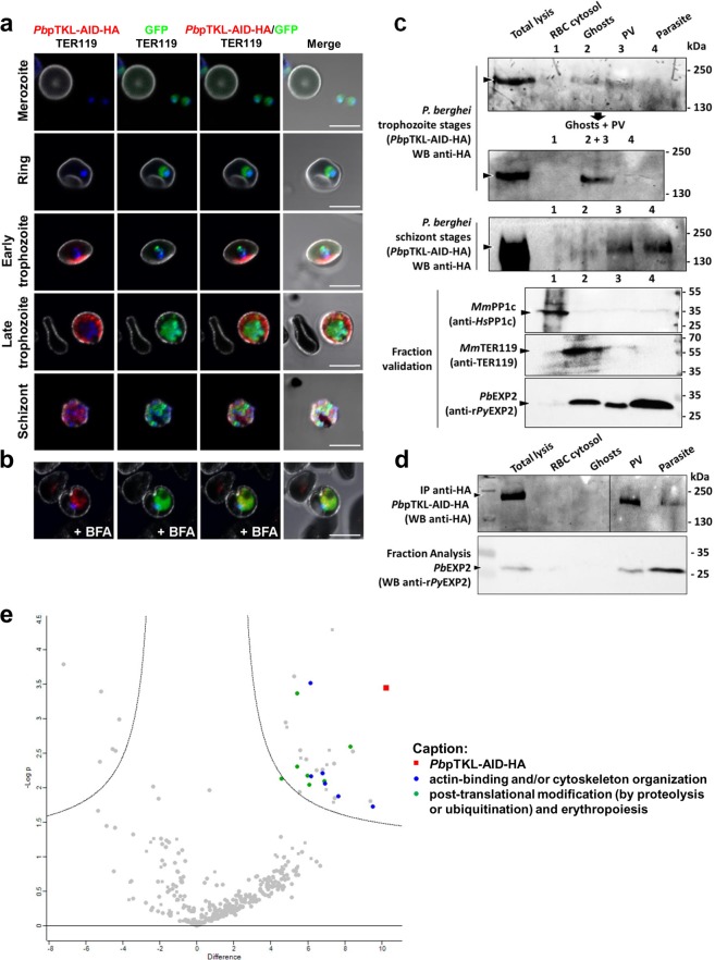
PbpTKL is exported in the erythrocyte during the trophozoite stages and binds host erythrocyte proteins. (a,b) PbpTKL is actively exported from the parasite between the trophozoite and schizont stages. (a) Confocal microscopy images showing the localization of the PbpTKL-AID-HA protein (red) in infected erythrocytes at different stages (top to bottom: merozoite, ring, early trophozoite, late trophozoite and schizonts). Parasite nuclei are stained with Hoechst (blue). The erythrocyte membrane is labeled after detection of TER119 (white). Bar = 5 µm. (b) Confocal microscopy images showing the localization of PbpTKL-AID-HA in brefeldin A (+BFA) treated trophozoites. PbpTKL-AID-HA (red, BFA aggregates) is no longer exported but retained within the parasite cytosol (GFP signal). Bar = 5 µm. (c) PbpTKL associates with ghosts during the trophozoite stages based on sequential lysis results (Supplementary Fig. S8). Fractions were verified by western blot (WB): anti-HsPP1c recognizes mouse PP1c in the erythrocyte cytosol, anti-TER119 recognizes mouse erythrocyte ghosts, and anti-rPyEXP2 recognizes PbEXP2 in the parasite cytosol, parasitophorous vacuole and vesicles in the host erythrocyte cytosol119. Anti-HA immunoprecipitation assays were performed on each fraction and PbpTKL-AID-HA was detected by anti-HA western blot. Uncropped blots are provided in the Supplementary Information. (d) PbpTKL is actively exported to the host erythrocyte cytosol. Sequential lysis on P. berghei trophozoites treated with BFA (verified by fraction analysis using anti-rPyEXP2, as opposed to fraction analysis without treatment shown in (c). Anti-HA IP assays were performed on each fraction and PbpTKL-AID-HA was detected by anti-HA western blot. Uncropped blots can be found in the Supplementary Information. (e) Exported PbpTKL binds erythrocyte proteins. Volcano plot showing proteins identified by mass spectrometry after anti-HA immunoprecipitation assays on PbpTKL-AID-HA versus wild-type pG230 P. berghei-infected erythrocytes (y-axis shows negative log p-value, x-axis shows fold-change between wild-type and PbpTKL-AID-HA samples). Squares = P. berghei proteins; circles = mouse proteins. Proteins absent from wild-type samples and detected in at least two of three PbpTKL-AID-HA ghost samples are shown on the upper right panel of the chart (see caption). Three biological replicates were analyzed per sample type (raw and interactome mouse protein analysis data are available in Supplementary Table S6).

