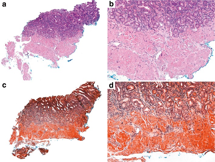
Figure 2.
Gastric biopsy. Parts (A) and (B) reflect hematoxylin and eosin-stained sections. Parts (C) and (D) reflect Congo red-stained sections. The gastric biopsy demonstrated pink amorphous material that showed apple-green birefringence on Congo red staining, which was consistent with amyloid deposition. For amyloid sub-typing, the specimen was sent for liquid chromatography tandem mass spectrometry, which showed a peptide profile consistent with AL (lambda)-type amyloid deposition.

