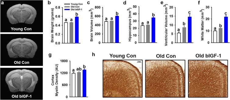
Fig. 3.
Phenotyping of young, old, and old bIGF-1 mouse brains by MRI and immunostaining. a–f Representative MRI brain images from young controls, old controls, and old bIGF-1 HET3 mice and corresponding results for weight and volume. Old control mice showed an increase in ventricular and white matter volume compared to young controls, while bIGF-1 mice showed an increase in brain weight and volume, hippocampal volume, ventricular volume, and white matter volume compared to other groups. Both males and females were assessed but are presented together due to similar patterns in outcomes [Young Control (n = 11 total; n = 6 females, n = 5 males), Old Control (n = 12 total; n = 5 females, n = 7 males), Old bIGF-1 (n = 12 total; n = 7 females, n = 5 males)]. g, h Results and representative images from myelin immunostaining in cortex showed no difference with age but an increase in myelin density was observed in old bIGF-1 mice compared to young controls [Young Control (n = 16 total; n = 8 females, n = 8 males), Old Control (n = 17 total; n = 9 females, n = 8 males), Old bIGF-1 (n = 13 total; n = 6 females, n = 7 males)]. Bars represent mean ± SE. Letters indicate a significant difference between groups, P ≤ 0.05

