Figure 2.
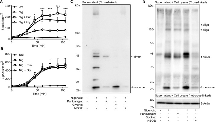
ASC speck release depends on cell lysis. A and B, LPS-primed (1 μg/ml, 2 h) ASC-mCherry iBMDMs were incubated with vehicle, punicalagin (Pun; 50 μm), or glycine (Gly; 5 mm) 15 min prior to activation with nigericin (Nig; 10 μm), and ASC speck formation was measured in real time without (A) or with (B) incubation of Z-VAD (50 μm) (n = 4). C and D, WT iBMDMs were treated as in A and B and activated with nigericin for 1 h. Supernatants (C) or combined supernatant and lysate (D) were cross-linked and analyzed for ASC or β-actin by Western blotting. ASC monomers (22 kDa), dimers (44 kDa), and oligomers are indicated. *, p < 0.05; ***, p < 0.001, determined by two-way ANOVA with Sidak's post hoc analysis and compared with the nigericin-treated group. Western blots are representative of three independent experiments. Error bars, S.D.

