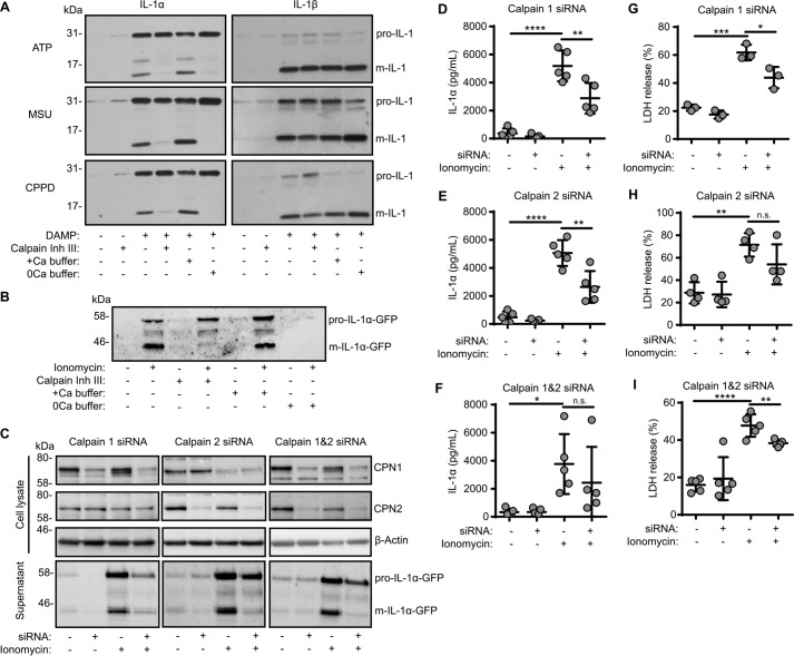
Figure 4.
IL-1α processing and release depends on calpains 1 and 2. A, LPS-primed (1 μg/ml, 2 h) peritoneal macrophages were incubated with calpain inhibitor III (50 μm) or in calcium-containing (+Ca) or calcium-free (0Ca) buffers 15 min prior to activation with ATP (5 mm, 1 h), MSU (250 μg/ml, 1 h), or CPPD (250 μg/ml, 1 h). Supernatants were analyzed for IL-1α (pro-form, 31 kDa; mature form, 17 kDa) and IL-1β (pro-form, 31 kDa; mature form, 17 kDa) by Western blotting. B, HeLa cells were transfected with pro-IL-1α-GFP (24 h) and then incubated with calpain inhibitor III (40 μm) or in calcium-containing (+Ca) or calcium-free (0Ca) buffers 15 min prior to activation with ionomycin (10 μm, 1 h). Supernatants were analyzed for IL-1α-GFP (pro-form, 58 kDa; mature form, 44 kDa) by Western blotting. C–I, HeLa cells were transfected with calpain 1, calpain 2, or scrambled siRNA (48 h), transfected with pro-IL-1α-GFP (24 h), and treated as in B. C, cell lysates were analyzed for calpain 1 (CPN1; 75 kDa), calpain 2 (CPN2; 75 kDa), and β-actin (42 kDa) and supernatants for m-IL-1α-GFP (44 kDa) by Western blotting. Supernatants were assayed for IL-1α release measured by ELISA (D–F) and cell death, measured as LDH release (n = 4) (G–I). *, p < 0.05; **, p < 0.01; ***, p < 0.001; ****, p < 0.0001; n.s., nonsignificant, determined by one-way ANOVA with Dunnet's post hoc analysis compared with the ionomycin-treated group. Western blots are representative of at least three independent experiments.

