Figure 7.
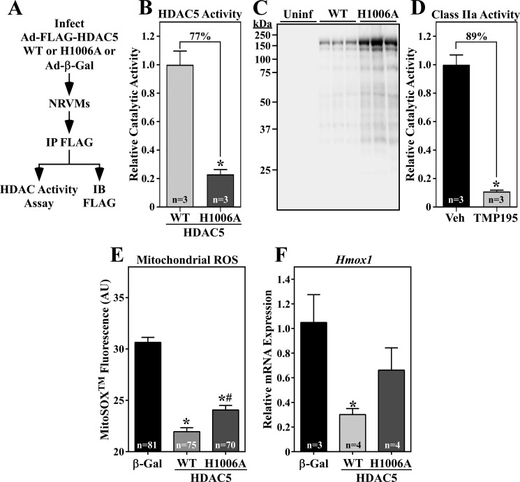
HDAC5 gain-of-function reduces cardiomyocyte oxidative stress and NRF2 target gene expression. A, schematic of the HDAC5 activity assay. IB, immunoblot. B and C, 72 h after infection, NRVM protein homogenates were immunoprecipitated with anti-FLAG antibody, and immunoprecipitates were incorporated into in vitro HDAC activity assays employing a class IIa HDAC–specific substrate, as described under “Experimental procedures.” A portion of each immunoprecipitate was subjected to immunoblotting with anti-FLAG antibody. Values represent means +S.E., with n = plates of cells per condition. The signal from immunoprecipitates of lysates from Ad-β-gal–infected NRVMs was subtracted as background, and the values were normalized to the amount of immunoprecipitated WT or H1006A determined in (C). *, p < 0.05 versus WT. Uninf, uninfected. D, class IIa HDAC activity was quantified in living NRVMs using a cell-permeable substrate. Values were obtained 5.5 h after treatment with TMP195 (3 μm) or DMSO vehicle control. Values represent means + S.E., with n = the number of independent wells of NRVMs quantified. *, p < 0.05 versus vehicle control. E, NRVMs were infected with an adenovirus encoding β-gal, WT HDAC5, or HDAC5 (H1006A) for 72 h. The cells were subsequently washed with PBS and incubated with MitoSOXTM (3 μm) in PBS for 20 min at 37 °C. Mitochondrial ROS levels were determined by live-cell imaging and quantifying the fluorescence intensity of MitoSOXTM in single cells. Values represent means + S.E., with n = the number of cells quantified. *, p < 0.05 versus β-gal; #, p < 0.5 versus WT. F, NRVMs were infected with adenoviruses as described above, and Hmox-1 mRNA expression was determined by qRT-PCR. Values represent means + S.E., with n = the number of plates of cells per condition. *, p < 0.05 versus β-gal.

