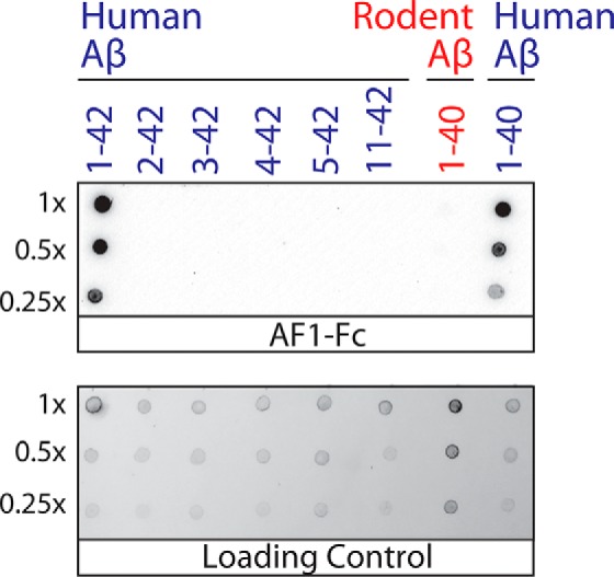
Figure 7.

Immunoblot analysis of the conformational epitope of the AF1 antibody. Human and rodent Aβ peptides and fragments thereof were assembled into thioflavin T-positive aggregates, purified via sedimentation, and deposited on nitrocellulose membranes. The samples were loaded at equal thioflavin T values for each dilution, except for human Aβ(1–40) fibrils (which had approximately five times lower thioflavin T values). AF1–Fc immunostaining was evaluated at 10 nm (1% milk). The loading control blot was detected using silver stain.
