
Fig. 1.
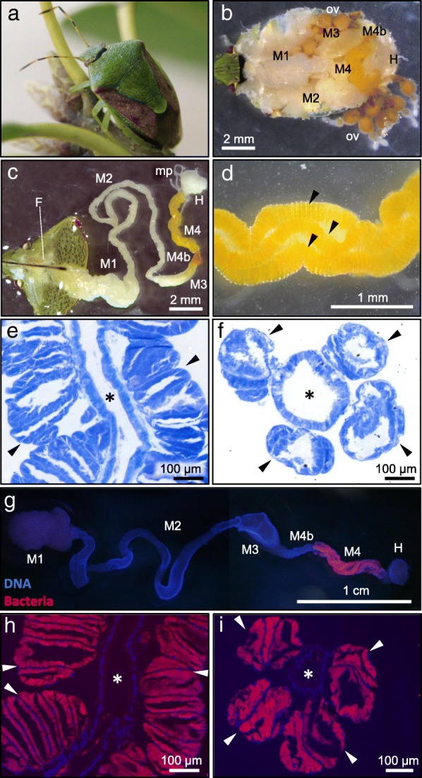
Symbiotic organ of P. stali. a An adult insect. b Dorsal view of a dissected insect. c An isolated alimentary tract, in which M4 represents the symbiotic organ. d An enlarged image of the symbiotic organ with numerous crypts in four rows. Three rows are seen (arrowheads), whereas another row is behind. e, f Toluidine blue staining of a longitudinal section (e) and a cross section (f) of the symbiotic organ. g FISH detection of the symbiotic bacteria in a dissected alimentary tract. h, i FISH detection of the symbiotic bacteria on a longitudinal section (h) and a cross section (i) of the symbiotic organ. In g–i, blue and red signals indicate host’s nuclear DNA and symbiotic bacteria, respectively. Arrowheads and asterisks show the crypt rows and the main tract of the midgut symbiotic organ, respectively. Abbreviations: F, foregut; M1, midgut first section; M2, midgut second section; M3, midgut third section; M4b, bulb-like section aboral to M4; M4, midgut fourth section, the symbiotic organ with crypts; H, hindgut; ov, ovary; mp, Malpighian tubule