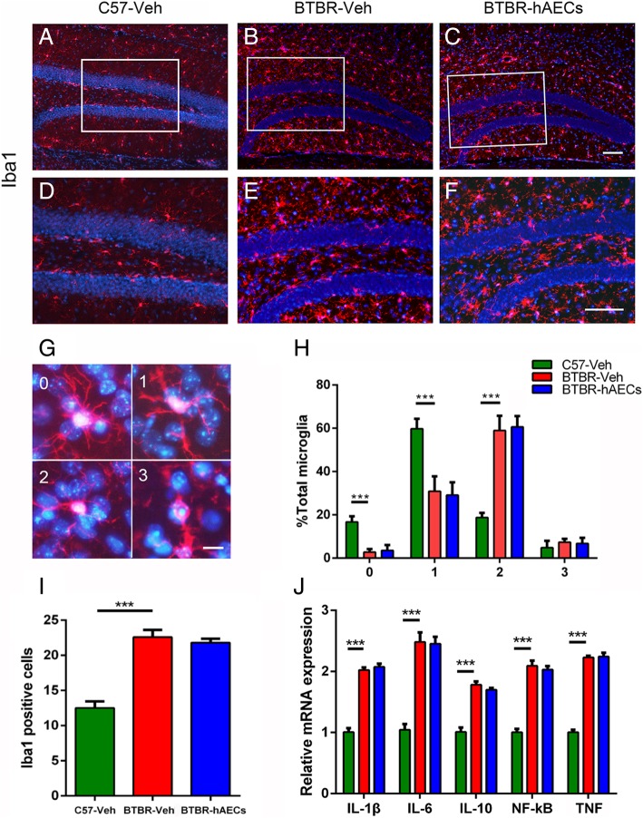
Fig. 3.
hAEC treatment did not alter microglia activation and inflammatory factors in the hippocampus of BTBR mice. a–c Microglia cells labeled by Iba1 in the DGs. d–f Magnified views of the boxed areas in a–c. g Microglia cells labeled by Iba1 of each category. h Quantitative analysis of the percentage of Iba1+ cells of each category. i Quantitative analysis of the number of Iba1+ cells in the SGZ. j In the hippocampus, the relative mRNA expression levels of IL-1β, IL-6, IL-10, TNF, and NF-κB, which are inflammatory cytokines, were expressed in high levels in BTBR mice with vehicle treatment compared to C57 mice; these expression levels were the same in the vehicle-treated and hAEC-treated BTBR mice. The data are presented as the mean ± SEM (n = 4 for immunohistochemistry, n = 3–4 for RT-qPCR). Scale bar in c = 50 μm and applies to a–c; in f = 50 μm and applies to d–f; in g = 20 μm

