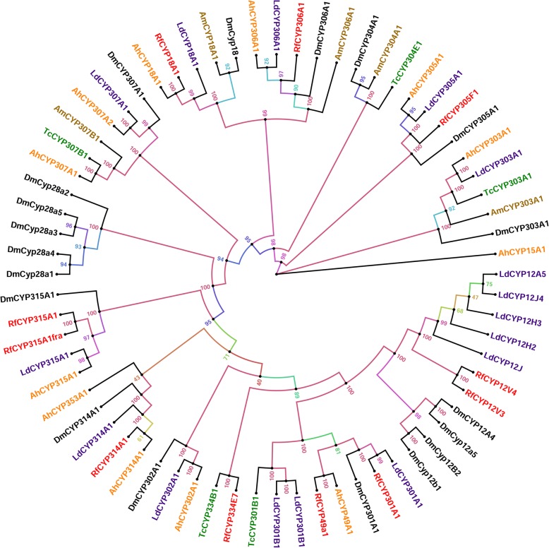
Fig. 3.
Maximum likelihood consensus tree of CYP2 and Mitochondrial clans. The phylogenetic tree reconstruction was performed using the same methods mentioned in Fig. 1, with LG + I + G + F substitution model determined as the best-fit model of protein evolution. P450s from Tribolium castaneum, Bemisia tabaci, Leptinotarsa decemlineata and Drosophila melanogaster P450s were used in the analysis to classify R. ferrugineus CYP2 and Mitochondrial clans. Additionally, candidates from Agasicles hygrophila and Apis mellifera were added for classification purposes. P450s from different species were marked with different colors. The phylogenetic tree was visualized using FigTree (http://tree.bio.ed.ac.uk/software/figtree/) and branch appearance was colored based on the bootstrap values. Scale = 2.0 amino acid substitutions per site

