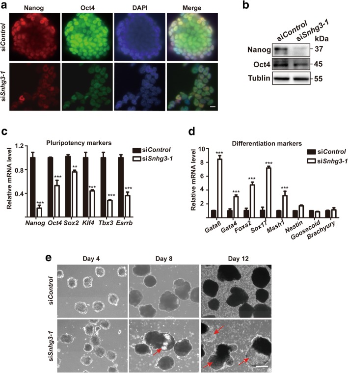
Fig. 3.
Snhg3 depletion resulted in mouse embryonic stem cell (mESC) differentiation. Immunostaining (a) and western blot (b) of mESCs after siControl treatment or siSnhg3 depletion to detect Nanog and Oct4 expression. The nuclei were stained with DAPI; × 20 objective, scale bar = 100 μm. Tublin was used as a loading control. qRT-PCR was used to analyze the expression levels of pluripotency markers (c) or differentiation markers (d) after siControl treatment or siSnhg3 knockdown in mESCs. Data are presented as mean ± SD; n = 3, two-way ANOVA. **p < 0.01; ***p < 0.001. e siControl- or siSnhg3-treated mESCs were cultured as hanging drops in ES medium without LIF to form embryoid bodies (EBs) for the indicated times. Red arrows indicate cystic EB formation; × 4 objective, scale bar = 400 μm

