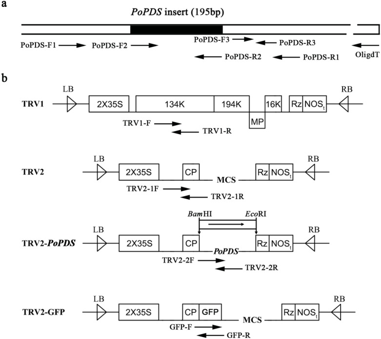
Figure 2. Schematic representation of TRV constructs used in this study.

(A) The cDNA of PoPDS insert for its introduction into TRV vector. PoPDS-F1/PoPDS-R1 was used to amplify the open reading frame region of PoPDS, PoPDS-F2/PoPDS-R2 targeted the inserted fragment ofPoPDS (the black box), and PoPDS-F3/PoPDS-R3 was designed for quantitative real-time PCR. (B) The structures of TRV1, TRV2, TRV2-PoPDS, and TRV2-GFP. The arrows indicate the different primer pairs for examining TRV1, TRV2-1, TRV2-2, and GFP transcript levels. LB left border, RB right border, MP movement protein, 16K 16 Kd protein, Rz self-cleaving ribozyme, NOSt NOS terminator, CP coat protein, MCS multiple cloning site.
