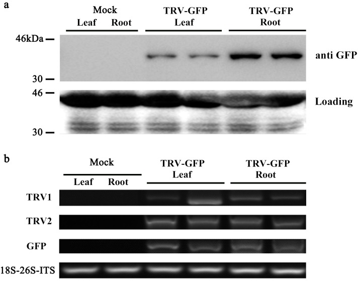
Figure 5. Detection of GFP protein accumulation in TRV-GFP-inoculated P. ostii leaves and roots.
(A) Western blot analysis of CP-GFP protein levels in mock treated, TRV-GFP-infected P. ostii leaves and roots at 5 days post inoculation. Ten micrograms of protein were loaded into each lane and an anti-GFP antibody was used to detect the CP-GFP fusion protein. Coomassie blue staining was used to confirm equal loading in each lane. (B) Semi-quantitative RT-PCR analysis of TRV1, TRV2, and GFP accumulation levels in mock treated, TRV-GFP-infected P. ostii leaves and roots. 18S-26S internal transcribed spacer (18S-26S-ITS) was used as internal control.

