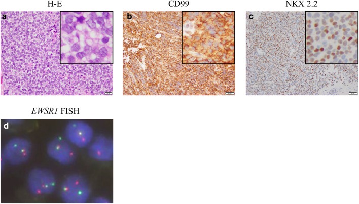
Fig. 1.
Histological and fluorescence in situ hybridization (FISH) findings of the tumor at diagnosis. Boxed images in the right upper corner are the magnification of each image. Hematoxylin–eosin (H–E) stain showed small round tumor cells (a). Immunohistochemical analysis demonstrated positivity for CD99 and NKX 2.2 (b, c). FISH demonstrated EWSR1 split signal pattern with a pair of fused yellow signals and split red (5′ EWSR1) and green (3′ EWSR1) signals (d)

