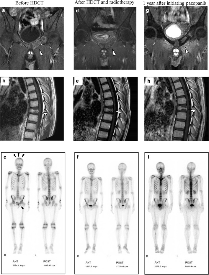
Fig. 3.
Contrast-enhanced T1-weighted magnetic resonance imaging and bone scintigraphy before the high-dose chemotherapy (HDCT) (a–c), after HDCT and radiotherapy (d–f), and 1 year after initiating pazopanib (g–i). Abnormal accumulations were noted in the cranial bone and pubis before HDCT (c). Abnormal accumulations in bone scintigraphy have diminished after HDCT and radiotherapy (f) and remained undetectable 1 year after initiating pazopanib (i). Lesions in the pubis have reduced in size, with almost negligible contrast enhancement during the treatment (a, d, and g). Remaining contrast enhancement in the thoracic vertebrae before HDCT (b) has diminished after HDCT and radiotherapy (e) and remained undetected 1 year after initiating pazopanib (h). Arrowheads indicate lesions (a, b, d, e, g, and h) and abnormal accumulations (c)

