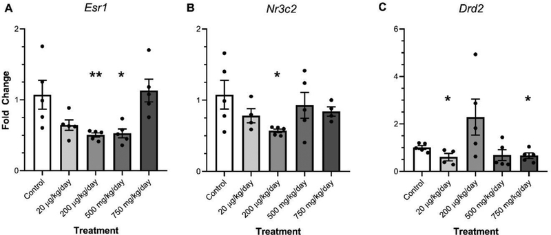
Figure 3.
The effect of ancestral DEHP exposure on gene expression in the amygdala of F3 females. The relative expression of Estrogen receptor 1 (Esr1) (A), Mineralocorticoid receptor (Nr3c2) (B), and dopamine receptor D2 (Drd2) (C) are shown for the F3 generation females. Data are representative of the means (± SEM). Control n=5, 4, and 4 respectively; 20 μg/kg/day n=5 for all; 200 μg/kg/day n=5, 5, and 4 respectively; 500 mg/kg/day n=5 for all; and 750 mg/kg/day n=5, 4, and 5 respectively for all analyses. **p<0.01, *p≤0.05 (significant difference compared with the control).

