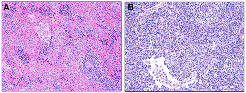
Fig. 4.

Representative histological images comparing H&E stained spleens from ETC and AP lambs. (A) ETC [H&E Stain]: this is a representative tissue control image with congestion/erythropoietic islands (score 3), EMH (score 3). and no necrosis, sinusoidal histiocytosis or pigmented macrophages (score 0); (B) AP [H&E Stain]: this is a representative image from AP lambs with sinusoidal histiocytosis (score 2), and no necrosis, congestion, pigmented macrophages, or EMH (score 0). ETC: Early Tissue Control; LTC: Late Tissue Control; AP: Artificial Placenta; EMH: Extramedullary Hematopoiesis.
