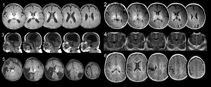
Figure 2.
Neuroimaging showing extent of lesion location in each participant. Images are multiple magnetic resonance imaging slices. Axial views are ordered inferior to superior, and sagittal views are ordered left to right. Infant 4 shows coronal views from cranial ultrasound. All axial images are in radiological orientation (top of image is anterior, left side of image is right side of the head). Participants’ corrected age at time of imaging for image sets 1–6 are 3 months, 3 months, 4 months, 0 weeks, 10 months, and 8 months, respectively.

