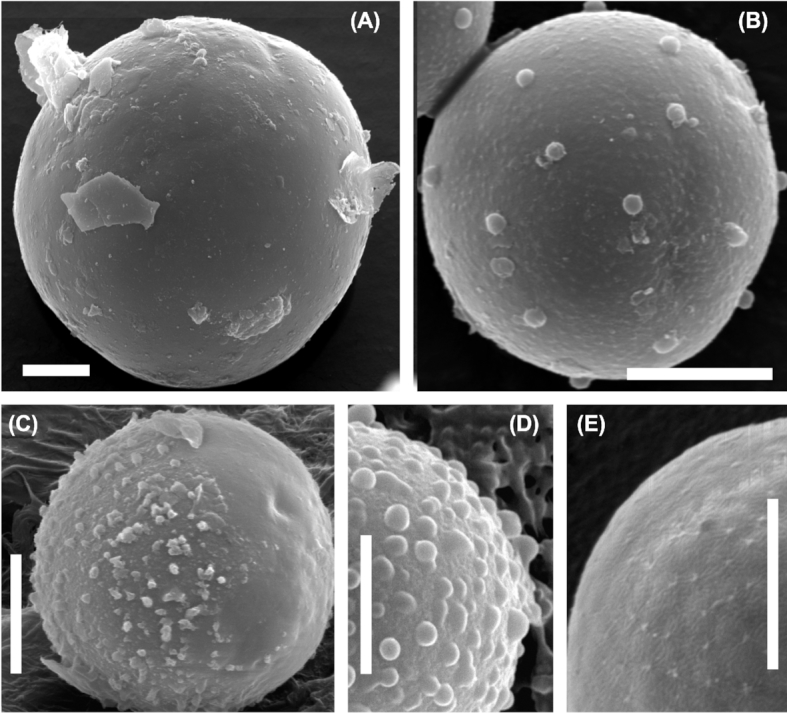
Figure 3.

Scanning electron microphotographs of Sanguina nivaloides. (A) Typical mature red cyst with smooth cell wall (holotype specimen RS_0015–2010). (B-D) Mature red cysts of less frequent haplotypes causing blooms, (B) showing nipples on cell surface (paratype specimen RS_0003–2004), (C) depicts the smooth surface with short blunt nibs, which may represent the compressed remnants of the presumably transient primary cell wall, this cell wall layer may decompose over time (note: on the left side of the cell 'papillae' are present, on the right side they are absent, specimen RS_0011), (D) shows the pronounced papillae on the cell surface (sample 4D). (E) Scattered cells with small papillae on their cell surfaces in a population of smooth-walled cells (sample redCol). Scale bars 5 µm.
