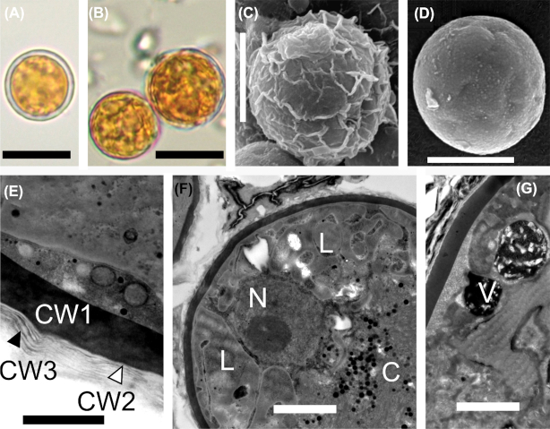
Figure 5.
Light and electron micrographs of Sanguina aurantia (holotype specimen RS_0017–2010), (A) depicts the distant outer cell layer. (B) shows that the distant cell layer is absent. (C-D) outermost layer is wrinkled and when it is decomposed, the smooth cell wall below may appear. (E) shows details of the cell wall with the electron-dense layer (CW1) being covered by several more or less undulating thin transparent layers (CW2, CW3). (F) shows a cross-section of the cyst showing the nucleus and the centrally compressed chloroplast. (G) depicts the cytosolic electron-dense vacuoles filled with crystalline structures. C = chloroplast, CW1 = cell wall 1, CW2 = cell wall 2, CW = cell wall 3, L = lipid globules, N = nucleus, V = vacuole. Scale bars = 10 µm for (A, B), 5 µm for (C, D), 0.5 µm for (E) and 2 µm for (F, G).

