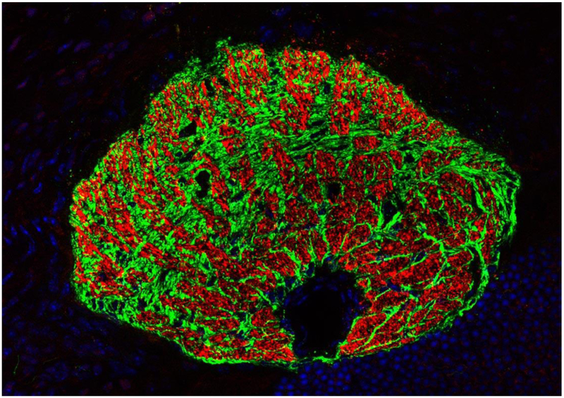
Figure 1. The glial lamina in the mouse eye.
Oblique cross section through the glial lamina shows that astrocytes (labeled in green) compartmentalize ganglion cell axons (labeled in red) into bundles forming glial tubes. The black area in the lower quadrant marks the position of the ophthalmic artery. Immunohistochemical staining for GFAP (green) and for neurofilament (red). Nuclei are labeled with DAPI (blue). Kindly provided by Rudolf Fuchshofer (University of Regensburg, Regensburg, Germany).

