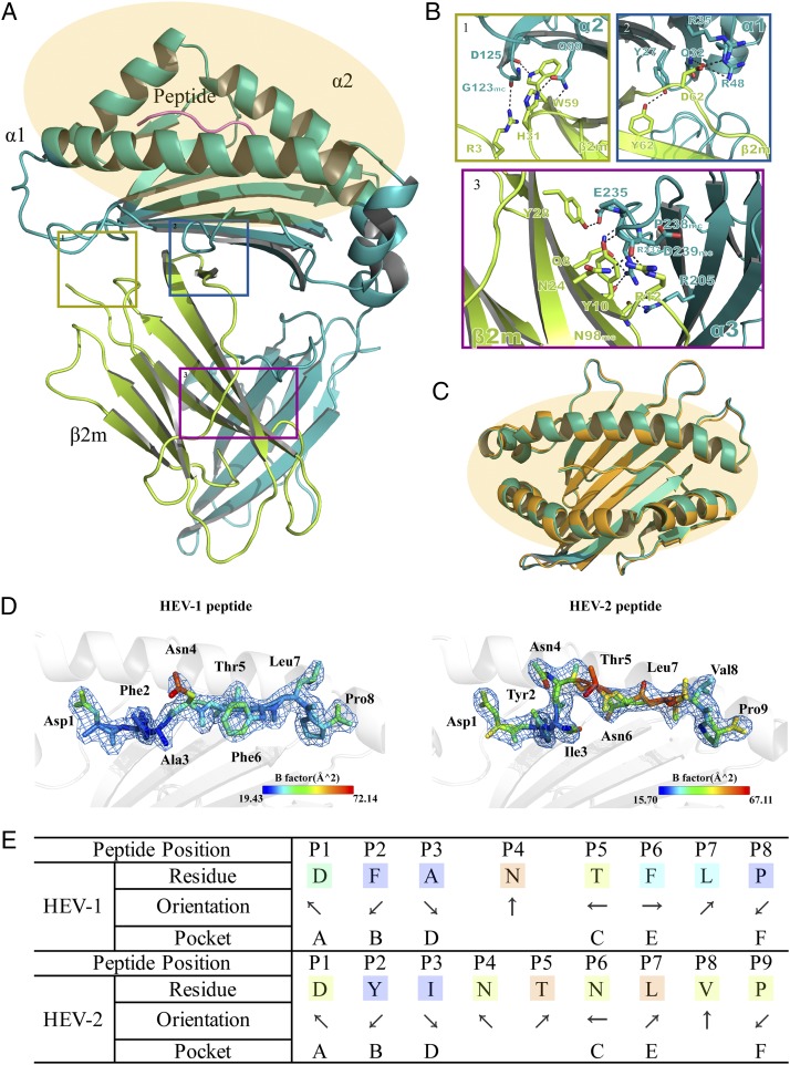
FIGURE 1.
Overview of p/Ptal-N*01:01/HEV-1 and binding peptide comparison with p/Ptal-N*01:01/HEV-2. (A) The HC of p/Ptal-N*01:01/HEV-1 is shown in teal, composed of three domains named α1, α2, and α3. The L chain, β2m, is shown in yellow. The peptide HEV-1 (DFANTFLP) is shown in violet. The ABG is highlighted in orange. (B) The H-bonds between β2m and the HC (from 1 to 3 showing the interactions in domains α2, α1, and α3, respectively). The H-bonds are represented by black dotted lines. The corresponding residues are labeled with amino acid abbreviations and primary sequence numbers. Mc indicates the main chain of the corresponding amino acids. Nitrogen atoms are shown in blue and oxygen atoms in red. (C) Comparison of the binding clefts of p/Ptal-N*01:01/HEV-1 (octapeptide) in teal and p/Ptal-N*01:01/HEV-2 (nonapeptide) in bright orange. With p/Ptal-N*01:01/HEV-1 as the reference, the RMSD between the HC was 0.436 Å, between β2m was 0.292 Å, and between the ligands was 1.814 Å. (D) The peptide comparison between HEV-1 and HEV-2 binding with p/Ptal-N*01:01 shown as a cartoon representation. Corresponding amino acids are labeled and colored by B-factor. (E) The orientation and location of HEV-1 and HEV-2 binding in the pocket of p/Ptal-N*01:01. The arrows pointing upward or downward represent the direction of the amino acid side chains toward the TCR or the MHC I base, respectively. The arrows pointing left or right represent the amino acid side chains facing the α1 domain or the α2 domain, respectively.

