
Figure EV3. Impact of actin filament growth on the number of microtubule nucleation sites in isolated centrosomes.
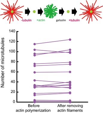
These data are associated with the set of experiments shown in Fig 4D–F and aimed at testing the impact of actin filament growth on the composition and structure of isolated centrosomes. Top line shows a schematic illustration of the dynamic assay: tubulin is added to measure centrosome nucleation capacity and washed out. Then, actin is added and later disassembled with gelsolin. This step of actin assembly/disassembly is followed by the addition of tubulin to measure again centrosome nucleation capacity. The amount of microtubule per centrosome is compared between the first and the last step of tubulin addition. No significant difference could be detected (Student's paired t‐test).