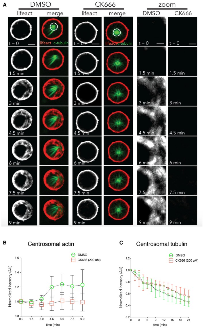
Figure EV5. Arp2/3‐dependent actin nucleation around centrosomes during monopolar exit.

- Representative image of cells arrested in prometaphase with STLC incubated for 2 h with DMSO or 0.2 mM CK666 was imaged every 90 s as they were forced to exit mitosis as the result of RO‐3306 addition. n = 26 cells (DMSO) and 27 cells (CK666) from two independent experiments. Scale bar = 5 μm and for zoom = 2 μm.
- Quantification of actin around centrosome for cells treated with DMSO or 0.2 mM CK666 prior to forced exit shows that pre‐treatment with 0.2 mM CK666 leads to a failure to accumulate actin around the centrosome during exit. n = 26 cells (DMSO) and 27 cells (CK666) from two independent experiments. Error bars indicated standard deviation.
- Quantification of tubulin around centrosomes for cells treated with DMSO or 0.2 mM CK666 prior to forced exit shows that pre‐treatment with 0.2 mM CK666 leads to a failure to decrease tubulin around the centrosome during exit. n = 26 cells (DMSO) and 27 cells (CK666) from two independent experiments. Error bars indicated standard deviation.
