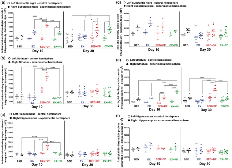
Figure 5.
Iba-1 immunostaining of activated microglia 10 days and 30 days after the surgery in the experimental (right) and control (left) substantia nigra (a), striatum (b) and hippocampus (c). Note the peak of microglial activation in the SED + PD animals at 10 days after the surgery for all the structures analyzed. At day 30, both groups of PD rats still revealed an increase in the microglial activation in the substantia nigra. Statistical differences between groups for the same brain hemisphere and between hemispheres for same group are presented as *p < 0.05; **p < 0.01; ***p < 0.001; ****p < 0.0001.
GFAP immunostaining of astrocytes 10 days and 30 days after the surgery in the experimental (right) and control (left) substantia nigra (d), in the striatum (e) and in the hippocampus (f). Note the peak of astrocyte activation in the striatum of PD rats at 10 days after the surgery, which normalized to control levels at 30 days in the exercised PD rats. Statistical differences between groups for the same brain hemisphere and between hemispheres for same group are presented as *p < 0.05; **p < 0.01; ***p < 0.001; ****p < 0.0001.

