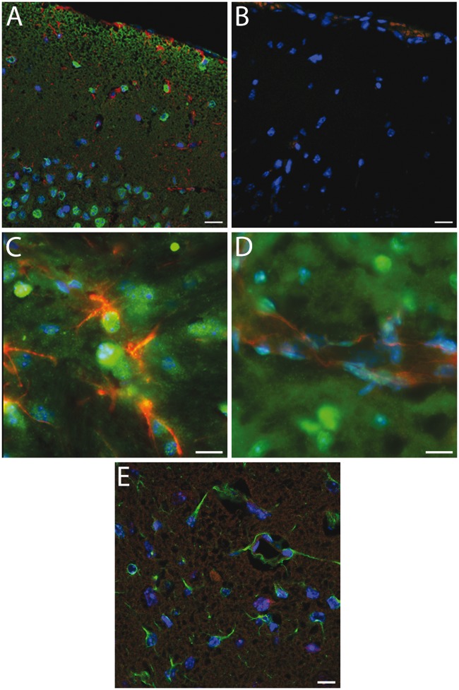
Figure 3.
Immunohistochemical labelling of Sgk1+/+ mouse cerebral cortex.(a) Robust cellular labelling for Sgk1 (green) is evident in cerebral cortical tissue from an unlesioned mouse. Cells within the cortical pyramidal layer are clearly labelled. There is relatively little overlap with the astroglial marker, GFAP (red). (b) As a negative control, a neighbouring section treated identically except for omission of primary antibodies, shows little non-specific labelling. (c, d) Higher magnification images confirm little overlap of Sgk1 labelling (green) with the astrocyte marker GFAP (panel C, red) or with an endothelial cell marker, CD31 (d, red). (e) Cortical tissue in a mouse at 48 h after MCAo. Ipsilateral cortical tissue within the ischaemic lesion. Astrocytic cells labelled with GFAP (green) are evident within the lesional area. Sgk1 labelling (red) is sparse. In all panels, nuclear chromatin is labelled with DAPI (blue). Scale bars 20 µm (a–b, e) or 10 µm (c–d).

