Figure 6.
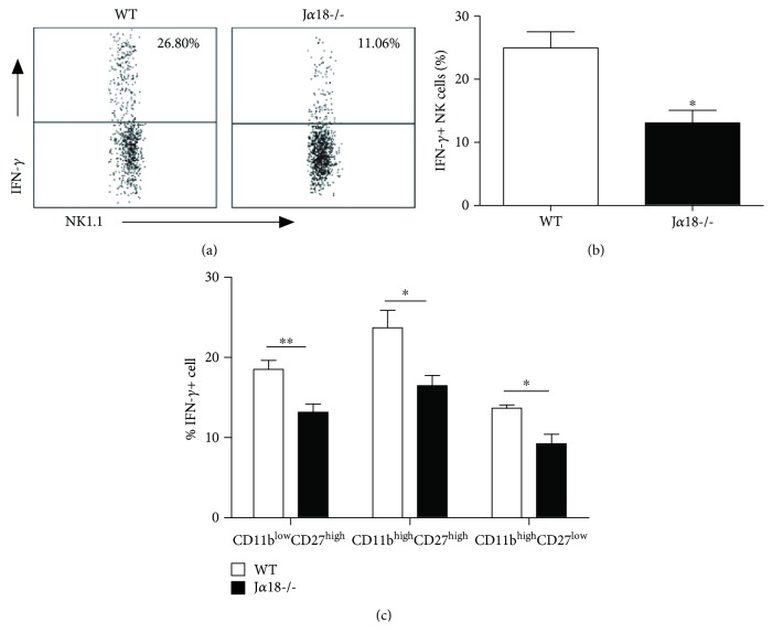
Reduced IFN-γ production by NK cells in Jα18-/- mice following Cpn lung infection. Mice were intranasally infected with Cpn (5 × 10⁶ IFU). At day 5 p.i., splenocytes from WT and Jα18-/- mice were stained for NK1.1, CD3e, CD11b, CD27, and IFN-γ and analyzed by flow cytometry. (a) Representative dot plots of IFN-γ production by NK cells in WT and Jα18-/- mice. (b) The percentage of IFN-γ-producing NK cells was summarized. (c) The frequency of IFN-γ production was analyzed on the specified NK cell subsets based on CD11b and CD27 staining. The data are shown as mean ± SD (n = 4). One representative experiment of three independent experiments is shown. ∗ p < 0.05.

