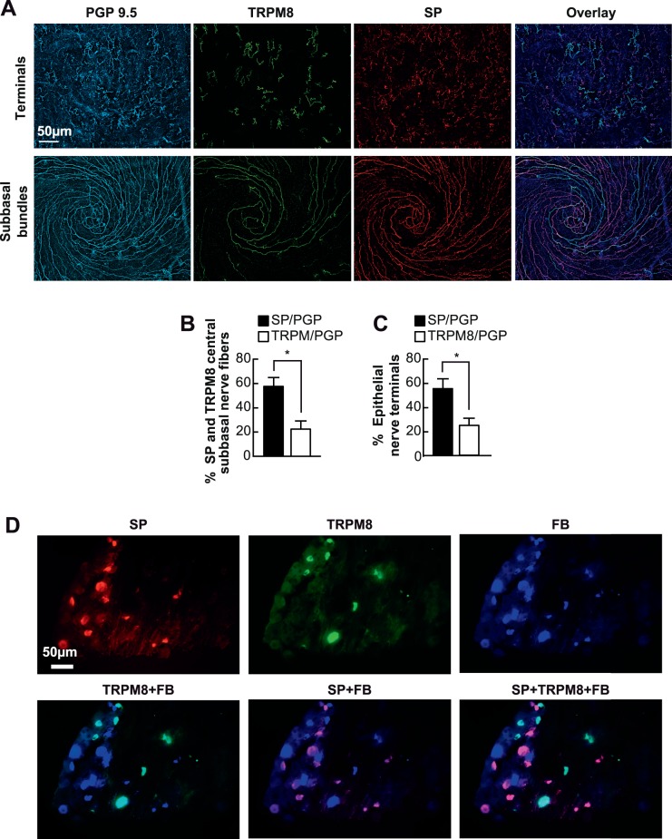
Figure 2.
Relative content of corneal epithelial TRPM8 and SP nerves. (A) Representative images of subbasal and nerve terminals in the vortex area labeled with PGP9.5, SP and TRPM8. (B, C) Percentage of TRPM8- and SP-positive subbasal bundles and nerve terminals versus total nerve area (PGP9.5-positive nerves) in each image. A total of 20 images for TRPM8 or SP and the same number of images for PGP9.5 were recorded from 10 corneas. Data expressed as average ± SD. (D) Localization of TRPM8- and SP-positive neurons in TG. Ipsilateral TG were FB-labeled through retrograde tracing. One week later, the whole TGs were processed for immunofluorescence with SP and TRPM8 antibodies. Representative images were recorded with a 20× objective lens.

