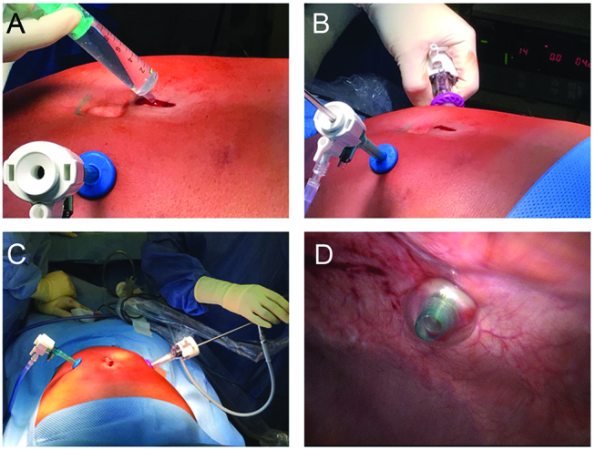
Figure 3:

Tightness of the minilaparotomy is verified by filling up the wound with saline, after insufflation of the abdomen (Panel A). After introduction under videoscopic control of a second trocar (Panel B), the intraabdominal position of the tip of the first trocar is visualized in order to exclude any bowel lesion (Panels C and D).
