Figure 1.
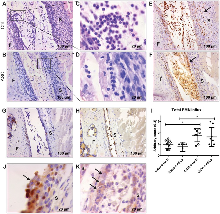
PMNs reallocate and cluster with ASCs in knees with early CiOA after intra-articular ASC injection. (A,B) Intra-articular injection on day 7 of CiOA resulted in attraction of immune cells within 6 h, both in the control (A) and ASC-injected (B) joints, as was shown in HE-stained total knee joint sections. (C,D) Higher magnifications showed that the immune cells in both saline- (C) and ASC-injected (D) CiOA knee joints had a polymorphonuclear (PMN) phenotype. (E,F) Immunohistochemistry with the specific antibody NIMP-R14 confirmed that a large number of PMNs is attracted to the joints. They were equally spread throughout the synovium and the joint cavity in the control-injected joints (E), but in the ASC-injected joints a remarkable accumulation of PMNs along the lining was found (F) as indicated by arrows. (G) NIMP-R14 staining showed that 24 h after ASC injection the PMN influx had largely disappeared. (H) ASC injection in naïve joints resulted in NIMP-R14 positive cell influx, albeit lower than in CiOA joints. (I) Quantification of attracted PMNs after intra-articular injection confirmed a significantly increased number of PMNs in CiOA joints compared to naïve joints. (J) At higher magnification we observed a clustering of PMNs around cells we supposed to be ASCs (arrow), which was confirmed with a CD271 antibody (K) which specifically stains ASCs (arrows). Control joints were CiOA knees injected with saline supplemented with 1% BSA. Images shown are representative for the treatment groups (n = 8 per group). Original magnification × 200 (A,B,E,F,G,H) or × 1,000 (C,D,J,K). Differences between groups were tested using a one-way ANOVA followed by a Bonferroni Multiple Comparison posttest. F, femur; S, synovium. Bars show mean values ± SD. *P < 0.05.

