Figure 2.
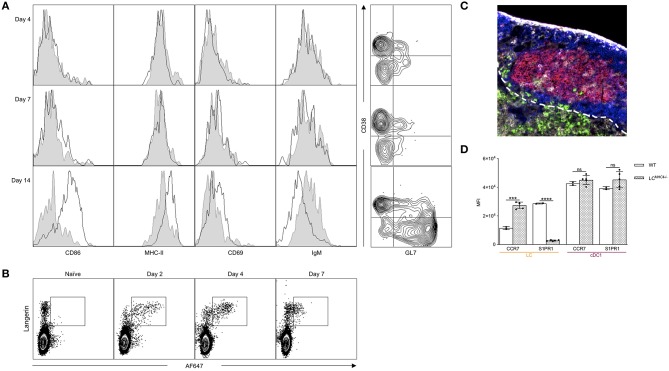
LCs can migrate to the B cell zone and activate B cells. (A) Left: Mice were immunized through LCs and the hIgG4-specific B cell phenotype determined by flow cytometry at the indicated timepoints. Black lines indicate immunized mice, while gray shaded histograms are from naïve mice. Right: The phenotype of the hIgG4-specific B cells displayed using CD38 and GL7 markers. One representative experiment out of three is shown. (B) WT mice were injected with 10 μg of AF647-labeled 4C7 antibody. At the indicated timepoints, the AF647 signal in LN Langerin+ cells (LC and cDC1) was determined by flow cytometry. One representative experiment out of three is shown. (C) Localization of LCs 14 days after immunization in a Batf3−/− mouse (red: PNA, blue: B220, green: Langerin, and gray: CD4). One representative experiment out of three is shown. (D) CCR7 and S1PR1 expression in LCs and cDC1s in WT mice and in mice in which LCs lack MHC-II. Each dot represents an individual mouse. One representative experiment out two is shown. ***p < 0.001, ****p < 0.0001, ns = not significant.

