
Figure 4.
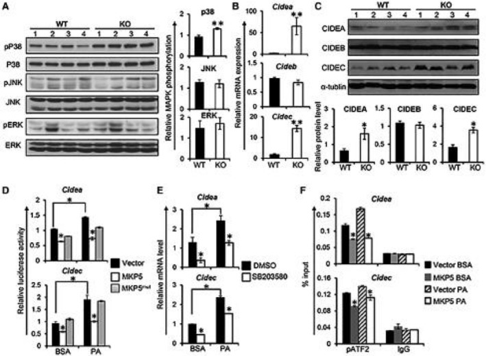
MKP5 regulates CIDEA and CIDEC/FSP27 in the liver through p38. (A) Representative western blots showing the activation of MAPKs, including p38, JNK, and ERK, in the liver from WT and MKP5 KO mice fed with the HFD for 12 weeks. Quantification of immunoblots was performed using ImageJ software. (B) mRNA expression of Cidea, Cideb, and Cidec/Fsp27 in the liver of WT and MKP5 KO mice following HFD feeding was determined by qPCR. (C) Protein levels of CIDEA, CIDEB, and CIDEC/FSP27 in the liver of WT and MKP5 KO mice were examined by western blots. (D) Overexpression of MKP5 inhibits Cidea and Cidec promoter activity. The Cidea or Cidec promoter region was cloned into a pGL3 luciferase vector and was separately cotransfected with MKP5 or MKP5mut together with a pRL reporter vector containing a cDNA encoding Renilla luciferase renilla luciferase‐null vector into FL83B hepatocytes for a dual‐luciferase assay after stimulation with or without PA. (E) p38 inhibition reduced the expression of Cidea or Cidec but not Cideb in mouse hepatocytes. FL83B mouse hepatocytes were pretreated with p38‐specific inhibitor SB203580 (40 μM) for 1 hour, followed by 500 μM of PA stimulation for 9 hours. Cide gene expression was examined by qPCR. (F) MKP5 attenuates pATF2 binding to the promoter of Cidea or Cidec/Fsp27. Mouse hepatocytes with or without MKP5 overexpression were treated with BSA or PA for 9 hours. Cells were harvested for the chromatin immunoprecipitation assay using pATF2 antibody to examine its binding to Cide promoters. The promoter region of Cidea or Cidec/Fsp27 associated with pATF2 was quantified by qPCR. Data shown are representative of three independent experiments with similar results. Data are presented as mean ± SEM. *P < 0.05; **P < 0.01. Abbreviations: BSA, bovine serum albumin; DMSO, dimethyl sulfoxide.