Figure 3.

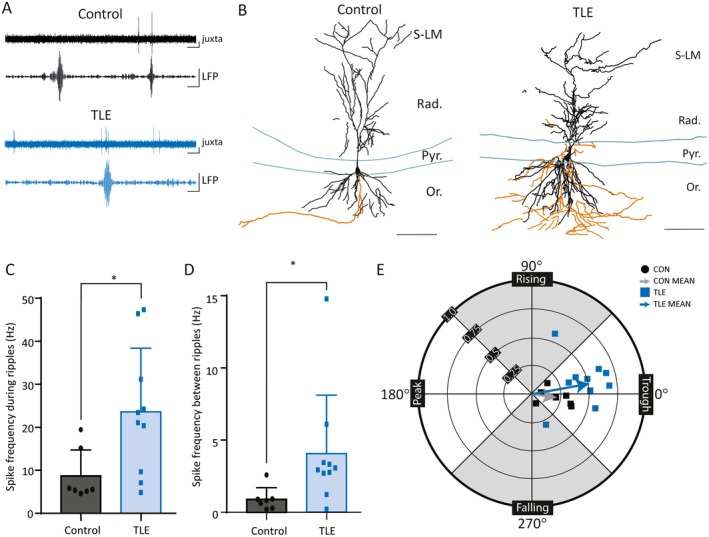
Increased firing of deep CA1 pyramidal cells during sharp‐wave ripples (SWRs) in temporal lobe epilepsy (TLE). A, Concurrent juxtacellular single‐cell activity (juxta) and local field potential network activity (LFP) demonstrating SWRs in control and epileptic mice. LFP traces filtered at 140‐250 Hz. Scale bars: 50 msec, 0.2 mV. B, Camera lucida reconstruction of a Neurobiotin‐filled deep pyramidal cell (dPC) (Vector Labs) in a control mouse (left) and in an epileptic mouse (right). Dendrites (black) and axon (orange) are reconstructed from three consecutive sections of 70 μm each. Layers of CA1 labeled as follows: Or, oriens; Pyr, pyramidal cell layer; Rad, radiatum; S‐LM, stratum lacunosum‐moleculare. Scale bars: 100 μm. C, D, Single cells exhibit increased firing frequency both during (C) and between (D) SWRs in epileptic mice when compared with control (* indicates P < 0.05). E, Wind rose plot in polar coordinates demonstrating the modulation strength (r) and preferred phase of firing (θ) of CA1 dPCs during SWRs in control and epileptic animals. The arrows are vectors representing the mean values. CA1 dPCs fire preferentially during the trough in both groups of animals, but the SWR exerts a higher degree of modulation on firing rate in the TLE animals
