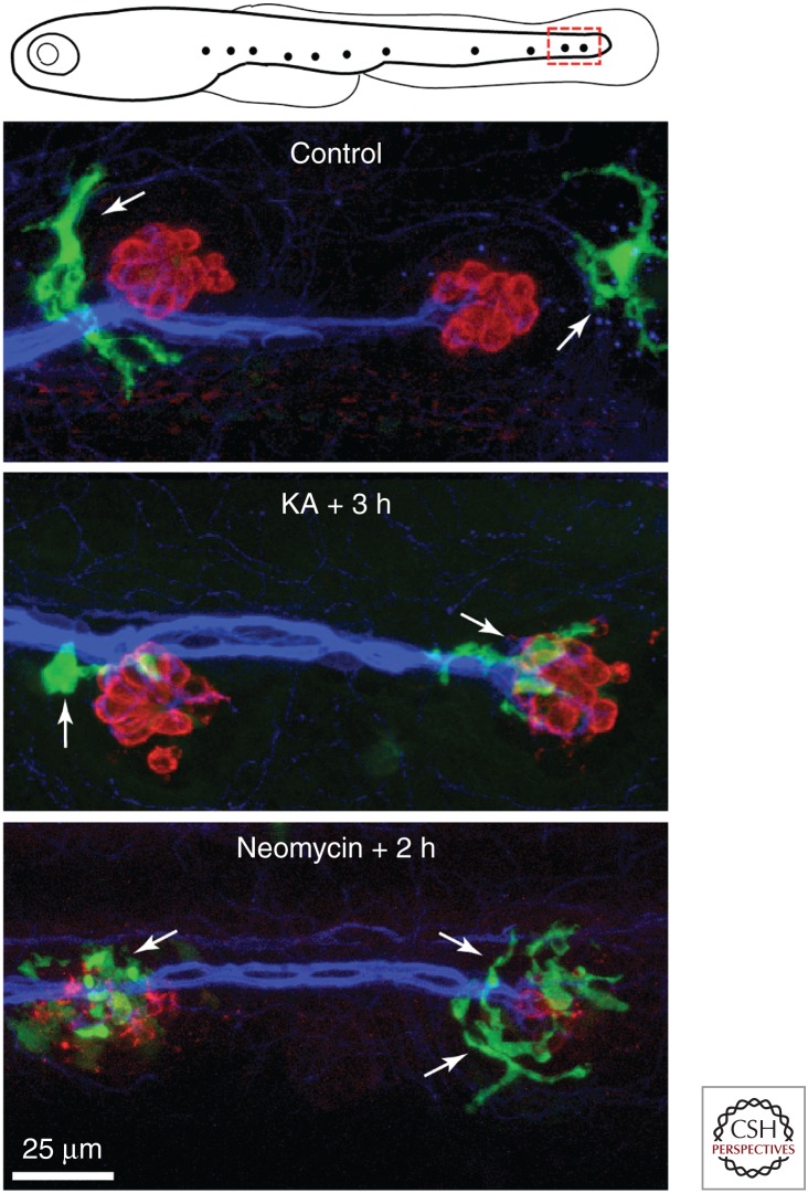
Figure 3.
Macrophage interactions with lateral line neuromasts in larval zebrafish. Transgenic fish lines in which macrophages express fluorescent proteins (e.g., mpeg1: green fluorescent protein [GFP]) permit the observation of macrophage activity in living fish. Published data show that neuromasts of the posterior lateral line typically possess one to two macrophages within a 50 µm radius of each individual neuromast (Hirose et al. 2017). Images show macrophages near the two most-posterior neuromasts of the zebrafish lateral line at 5 days postfertilization (see red box in top schematic). Macrophages are commonly observed near neuromasts in fish with intact hair cells (Control). However, injury to neuromasts by application of kainic acid ([KA], 300 µm for 1 h) or neomycin (50 µm for 30 min) causes macrophages to enter neuromasts and contact both injured hair cells and their neurons. Labels: green (GFP, macrophages), red (HCS-1, hair cells), blue (ZN-12, afferent neurons).

