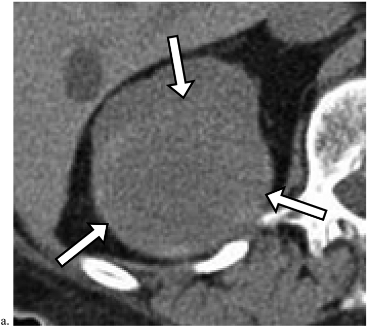
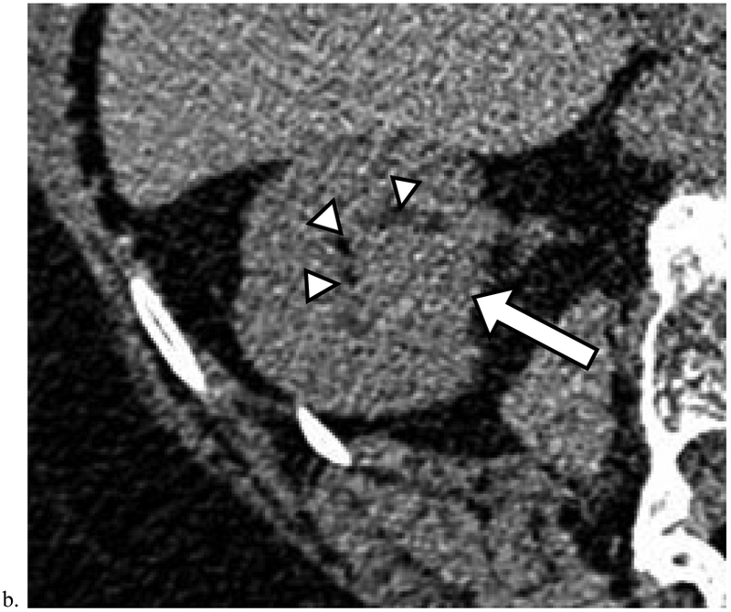
Figure 4. Missed Renal Cancers that Progressed Prior to Diagnosis.


a). Heterogeneous masses which deform the renal contour may represent renal cancer. Axial unenhanced CT image obtained in a 74-year-old male shows a 7.0 cm heterogeneous mass in the right kidney (arrows). Although this finding was identified, no diagnosis or follow-up recommendation was offered in the radiology report. The mass was diagnosed 8 months later as renal cell carcinoma (RCC) (Table 5, row 3). The patient died of non-small cell lung cancer one year later. b). Deformity of the inner renal contour aids in detection of renal cancer. Axial unenhanced CT image obtained in a 78-year-old female shows a 2.5 cm homogeneous mass (arrow) measuring 40 HU deforming the inner renal contour and displacing the renal sinus fat (arrowheads). This finding was not included in the original radiology report. The mass was diagnosed 46 months later as RCC (Table 5, row 1).
