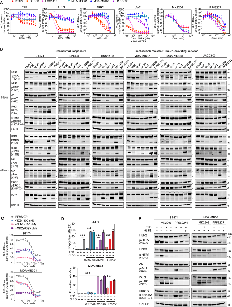
Fig. 4. FAK1 activation in response to AKT inhibition and effects of combination treatments.

(A) XTT cell proliferation assays of HER2-positive breast cancer cell lines with or without PIK3CA point mutations [BT474 (K111N), SKBR3 (WT), HCC1419 (WT), MDA-MB361 (E345K), MDAMB453 (H1047R) and UACCC893 (H1047R)] treated for 4 days with the indicated concentration of HER2-targeted therapy (TZB, 6L1G, ARRY, A+T), AKT inhibitor (MK2206), or FAK inhibitor (PF562271). Data are mean ± SD of n=3 experiments. (B) Western blot analysis of a HER2-dependent signaling cascade after 6 (top) and 48 hours (bottom) of treatment with the indicated drug (DMSO, 100 nM TZB, 100 nM 6L1G, 10 μM ARRY380, 10 μM PF562271, or 5 μM MK2206) or combination (A+T, 10 μM ARRY380 and 100 nM TZB). Blots are representative of 2–4 independent experiments. (C) XTT cell proliferation assays of BT474 and MDAMB361 cells after 4 days of continuous treatment with titration of the FAK1-inhibitor PF562271, and subsequent addition at day 0 (less than 5 min) of the indicated anti-HER2 agents TZB (100 nM) and 6L1G (100 nM), or the AKT-inhibitor MK2206 (5 μM). Data are mean ± SD of n=3 experiments. (D) High-throughput microscopy analysis of breast cancer cells continuously treated for 3 days 10 μM ARRY-380 or 10 μM PF562271 in combination with 100 nM TZB or 6L1G, then stained with propidium iodide (PI) and Hoechst-33342 to detect the number of membrane-permeable (PI-positive, inferred as dead) cells in the population. Data are mean ± SD of n=5 experiments. ***P ≤ 0.005 by one-sided pairwise t-test. (E) Western blot analysis for the indicated proteins in BT474 and MDA-MB361 cells treated for 2 days with 10 μM PF562271 or 5 μM MK2206, either alone or in combination with either 100 nM TZB or 6L1G. Blots are representative of 2 independent experiments.
