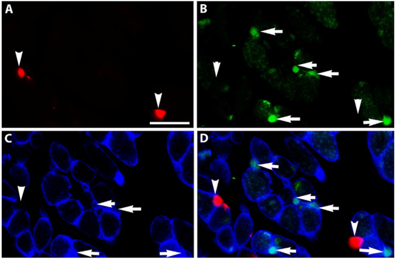
Fig. 3. RXRα does not co-express with Pax7.

Cryosection of an EOM from a Pax7-reporter mouse (red). The section was immunostained for (B) RXRα (green), (C) dystrophin (blue), and (D) is the merged image. Arrowheads indicate Pax7-positive cells and arrows indicate RXRα -positive cells. No co-expression was seen. Note there are also RXRα-positive nuclei (green) inside the sarcolemma. Scale bar is 20 μm.
