
Fig. 4. EECD34 cells maintain Pitx2 expression in vitro.
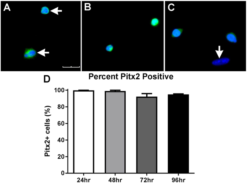
Representative images of EECD34 cells from wild type mouse EOM after (A) 24 h, (B) 48 h, or (C) 72 h in vitro in proliferation media immunostained for Pitx2 (green) and DAPI (blue). The bottom cell in (A) is dividing, and both are Pitx2-positive. Horizontal arrows indicate Pitx2 positive cells. Vertical arrow indicates a cell negative for Pitx2 (C). Scale bar is 30 μm. (D) Percentage of EECD34 cells from wild-type mouse EOM positive for Pitx2 after 24, 48, 72, and 96 h in vitro.